Tuesday 23 September 2008

Back Pain and Osteoporosis

Spinal fractures that occur as a result of osteoporosis are actually quite common, occurring in approximately 300,000 people in the UK each year. The problem is that the fracture is not always diagnosed instead, the problem is often just thought of as general back pain, such as from a muscle strain or other soft tissue injury, or as a common part of aging. Because of this, approximately two thirds or 200,000 of the vertebral fractures that occur each year are not diagnosed and therefore not treated.
Spinal fractures due to osteoporosis often occur while doing something that causes relatively minor trauma to the spine, such as opening a window, an insignificant fall, or twisting while lifting. Advanced cases of osteoporosis can even lead to a vertebral fracture with routine activities that would normally not cause any trauma, such as sneezing, coughing or turning over in bed.
Vertebral fractures are usually followed by acute back pain, and may lead to chronic pain, deformity (thoracic kyphosis, commonly referred to as a dowager's hump), loss of height, crowding of internal organs, and loss of muscle and aerobic conditioning due to lack of activities and exercise.
A combination of the above problems from vertebral fractures can also lead to changes in the individual’s self-image, which in turn can adversely affect self esteem and ability to carry on the activities of daily living.

It’s important to note that fractures from osteoporosis don’t just occur in the elderly, they can also occur in people as young as 40 or 50 years old. Because osteoporosis is a “silent” disease, meaning that there are typically no symptoms until a fracture occurs, it is not uncommon for someone with back pain to be unaware of the fact that she has actually fractured a vertebra (or multiple vertebrae) in her spine.

Osteoporosis is fairly common disease, and is especially common in postmenopausal women. In fact, in a recent article published in the USA it estimated that approximately 25 percent of all postmenopausal women in the United States have had a vertebral compression fracture.

Osteoporosis also occurs in men, but is far more prevalent in women approximately four times as many women have low bone mass, or osteoporosis, as men.
Osteoporosis causes bones to thin and become more brittle and weak. When the bones in the spine weaken they can break or cave in under normal pressure. The thinning bones can collapse during normal activity, leading to a spinal fracture. These compression fractures can cause a great deal of pain and can permanently alter the shape and strength of the spine.

The type of break in the spine that is typically caused by osteoporosis is called a compression fracture, usually defined as a vertebral bone in the spine that has decreased at least 15 to 20% in height due to fracture (as seen on an x-ray). These compression fractures can occur in vertebrae anywhere in the spine, but they tend to occur most commonly in the upper back (thoracic spine), particularly in the lower vertebrae of that section of the spine (e.g. T10, T11, T12). They rarely occur above the T7 level of the spine.

With a compression fracture caused by osteoporosis, the fracture usually occurs in the front of the vertebra, collapsing the bone in the front of the spine and leaving the back of the same bone unchanged. This results in a wedge shaped vertebra. Because the majority of damage is limited to the front of the vertebral column, the fracture is usually stable and rarely associated with any nerve or spinal cord damage.

Unlike many other conditions that can be treated before a serious complication develops, usually a fracture is the first sign that someone has osteoporosis. By the time a fracture occurs, the osteoporosis is usually advanced and the individual is then susceptible to more vertebral fractures.

Sunday 27 July 2008

Cauda Equina


Cauda Equina is known as a group of nerve roots at the lower end of the spinal cord.


What is Cauda Equina Syndrome?

The rare condition that affects the nerve endings at the bottom of the spinal column which can have severe consequences to a person’s mobility if it is not dealt with immediately is known as Cauda Equina Syndrome.
This condition weakens the nerves, which then receive electrical impulses sent from the brain that permit the lower limbs and pelvic organs to carry out they purpose properly. If this condition is not dealt with it can cause permanent paralysis, problems with the bowel and bladder and impotency.

This condition can occur if a person has suffered a viral infection, narrowing of the spinal canal or a slipped disc which has not been treated. Also many spinal injuries are bought on by a violent jolt or blow such as one might obtain if involved in a car accident. Spina Bifida is a congenital deformity (an abnormality in the closure of the spinal canal) which can also play a part.

Symptoms of Cauda Equina Syndrome
This condition has an extensive and altering mix of symptoms related to it but remember if any of the symptoms below are experienced then you should seek advice from your GP straight away.
• Stiffness when standing• Tingling prickling feeling in the legs, buttocks, thighs or feet • Numbness or pain in the legs• Bowel Dysfunction• Bladder weakness• Impotency
If any symptoms mentioned above appear over a continued period of time you should visit your doctor and give as much details as possible about the nature of the pain and the symptoms you have been experiencing.

In some cases the condition is only a temporary one that can be cured but for that to happen you have to give your doctor as much information as you can, that is very important. In these less serious cases the myelin covering that surrounds the nerves can become damaged so this can result in a temporary dysfunction, which can last quite a few weeks to a number of months.
In the more severe cases, the nerves (axons) be damaged but the good thing is that the nerves that run to the muscles (motor nerves) can grow back but this can be quite limited and can usually only be achieved after having surgery.

Diagnosis
Your doctor will examine you and assess your stability, reflexes, ability to stand and walk and also muscle strength. Your doctor may also ask for blood tests to be done and in severe cases a lumbar puncture too, which is when a needle is inserted into the lower spinal column to draw fluid for testing.

On top of this your doctor may also need x-rays, MRI (Magnetic Resonance Imaging) and CT (Computerised Tomography) scans to try and put together a more thorough picture.
Again it is extremely important that if you believe you have any of the symptoms mentioned that you contact your doctor at once so that he or she can take the proper steps to try and limit the damage. It is vital to remember that not only can this problem cause severe problems with the spine and the lower limbs but it can have unpleasant effects on the bowel, bladder, and sexual organs, which, if not treated, can lead to almost certain paralysis.

It is also very important to make sure that you continue sitting with the correct posture in the workplace and if it is necessary for you to lift any heavy objects then you do so in the proper way.

Back Trouble UK


Wednesday 23 July 2008

Rolfing

Rolfing was invented by Ida P. Rolf (1896-1979), who received her PhD in biochemistry and physiology from Columbia University in 1920. She went on to work at the Rockefeller Institute in the departments of chemotherapy and organic chemistry.
Finding the available therapy methods of her time inadequate, Dr. Rolf investigated the effects of structure on function in her search to find solutions to health-related problems. The catalyst to this search was a deal she made with a piano teacher that suffered from muscular dysfunction in the hands. Dr. Rolf wanted very much for her children to learn piano from this particular teacher. If she was able to improve and correct the teacher’s disability Dr. Rolf did improve the piano teacher’s disability and thus began to teach classes on Rolfing worldwide. In 1971 she established The Rolf Institute in Boulder Colorado. In addition, there are currently Rolfing centers in the United Kingdom, Europe, Asia, South America and Australia.


What Does ROLFING DO?
To see how Rolfing works is to look at the body in the gravitational field as a a fish in a body of water. As a fish is supported and lifted by water, we as humans can be supported and lifted by gravity. It is easy to see the lack of structural balance in an older person, bent over a cane. These imbalances don’t happen over night, they continually deteriorate the body’s capabilities. All of an individual’s history is recorded in their tissues, including falls, illnesses and psychological stress. Even the way a child copies a parent leaves a memory.


The human body’s adaptive nature will always work to integrate an injury. Rolfing helps release the body’s structure from the effects of time and trauma so that the body can do its job more effectively and efficiently. Preferably, Rolfing is used as preventive care, but when an injury is present it can enhance and expedite the healing process.


Who Can Benefit FROM ROLFING?
Rolfing can help anyone and everyone move closer to balance and vitality. The question is are you ready? In Rolfing, the client must actively participate in bringing their body back to balance in and out of the session. The session is only the beginning with increasing change continuing in the body as the client moves through their daily routine.


What Can I Expect in a ROLFING SESSION?
First of all, Rolfers are specialists in facilitating rapid and intense results. The goal is not pain instead its mechanisim is to melt the restrictions that are located at the origin and insertion points on the bone. With pressure and direction from the Rolfers hands and joint movement from the clients, synchronicity happens. Different tissues respond differently to pressure, the Rolfers job is to unlock the code for the different levels of tissure and fluide that surround the particular parts of the body. In some ways, like a hot iron press ironing out melting adheasions from dehydration, adheasions, lactic acid, injury, etc. Rolfers encourage rehydration and fluid consistancy at all spectrums of bodyily operations to support and maximize connection to the bodily fluids that promote regulartory functions for tissue health.


Clients can expect to move limbs and joints while the Rolfer with presssure, moves and holds certian tissues to create an energetic and yet physical release to allow tissues to resume a more healthy and balanced state. Every body is different so every body has it's own centerpoint, in respects to what bone alignment pattern is for them, balanced. The Rolfers job, is to help find what that is for the individual, their a guide of sorts, that facilitates growth at the mechanical level and for some also at the behavioral level. After all, there is the body, mind, spirit connection that is spoken in all tongue's, religons, cultures, myths, and legends.


To create a successful session, the client must be willing to explore new ways of operating in the body. This will help the client find a new and improved manner of movement and function that supports daily physical duties and personal expectations. This attitude will fuel the ability to interact effectively and efficiently in one’s own environment.


The Rolfing format is primarily an educational process and therefore implies an ongoing communication between the Rolfer and client. The client is often asked to make certain moves while the Rolfer is working. This helps the client to find and feel the new patterns of movement. The relationship between the Rolfer and client is a unique one. As trust, honesty and openness develop, the Rolfing process becomes an arena for personal transformation on the part of both individuals.

Tuesday 8 July 2008

(NMT) Neuromuscular Therapy

Neuromuscular Therapy (NMT) is a very specialised form of manual therapy. A therapist trained in NMT is educated in the physiology of the nervous system and its effect on the muscular and skeletal systems. The Neuromuscular Therapist is also educated in kinesiology and biomechanics and how to work in a clinical or medical environment.

By definition, Neuromuscular Therapy is the utilisation of static pressure on specific myofascial points to relieve pain. This technique manipulates the soft tissue of the body (muscles, tendons and connective tissue) to balance the central nervous system.

Neuromuscular Therapy will be used to address five elements that cause pain:
1.Ischemia: Lack of blood supply to soft tissues which causes hypersensitivity to touch
2.Trigger Points: Highly irritated points in muscles which refer pain to other parts of the body
3.Nerve Compression or Entrapment: Pressure on a nerve by soft tissue, cartilage or bone
4.Postural Distortion: Imbalance of the muscular system resulting from the movement of the body off the longitudinal and horizontal planes
5.Biomechanical Dysfunction: Imbalance of the musculoskeletal system resulting in faulty movement patterns (i.e., poor lifting habits, bad mechanics in a golf swing or tennis stroke or perhaps computer keyboarding) .

For a variety of reasons, when we strain muscles and fascia, either through impact or through a build up of chronic stress, the muscles go into spasm. This spasm can restrict blood flow and cause pain, as well as restricting mobility.
Sometimes muscles can be in tension for so long that they lack the energy to release, and form tight "knots" or "trigger points", a "contracture" or little hard bump in the muscle which can be responsible for debilitating back pain or neck & shoulder pain referring to other parts of the body. Fibromyalgia is a different condition, but it seems many patients with fibromyalgia have significant trigger point discomfort also.
Inflammation is a necessary part of the healing process, bringing nourishment into the area, and reducing flow away, which reduces the spread of infection. If allowed to continue and become chronic, however, it can lead to restrictive adhesions and thickening of connective tissue.
Neuromuscular Therapy involves releasing Trigger Points in muscles & fascia, and encouraging flexibility in muscle and connective tissue.
Pressure is applied to these Trigger Points, until the congestion diminishes. A release of tension relieves pain and increases mobility. Greater blood flow will allow the healing process to take over.
Stretching the muscle and fascia afterwards helps to consolidate the relaxation, and you will be shown some stretches to practice after the session.Your therapist should conduct a postural assessment to evaluate which areas to address.
Neuromuscular Therapy can be researched easily on the web, and it is gaining ground as a key modality for the repair of soft tissues.

Terry O'Brien

Tuesday 24 June 2008

Osteomyology

Our hectic lifestyles ask a lot of our bodies so it should come as no surprise when, our bodies protest!

An Osteomyologist may well be able to help you. Increasingly, they are becoming an indispensable part of an integrated medical service that complements, supports and augments traditional health care practice.

Although treatment is not yet available on the NHS, it is widely available throughout the U.K., with home visits and 24 hour emergency cover sometimes available. However, most treatments take place within the privacy of a clinic or private treatment room.

Osteomyology, breaks down to Osteo (bone) myo (muscle) ology (study of) so, taken literally, it is the study (and specialisation) of bones and muscles.

The application of this knowledge of the body gives rise to a wide range of physical therapy techniques which are utilised and refined constantly by the Osteomyologist.

Osteomyologists are often already qualified experts in their own field which may be Osteopathy, Chiropractics, Manipulative Science, Kinesiology, Physiotherapy or indeed any one of a number of manually based medical disciplines.

When the practitioner feels restricted by the constraints of his/her chosen field, and find they are lacking the tools to achieve a satisfactory improvement in their client, the natural progression is to widen their knowledge beyond the limitations of their society and training – This is the most common reason for joining the ever expanding ranks of Osteomyologists.

What can an Osteomyologist treat?
1. Joint pain and neurological symptoms.
2. Back and neck pain including whiplash.
3. Pregnancy related symptoms including sciatic pain.
4. Sports injuries.
5. Chronic conditions such as arthritis.
6. Repetitive strain injury.
7. Posture problems.
8. Musculoskeletal dysfunction including reduced flexibility of the joints and spine.


Treatment has also been shown to be effective for the following conditions:
1. Migraine and headaches.
2. Period pains.
3. Behaviour problems in children.
4. Diabetes.
5. Stress.
6. Asthma.
7. Glue ear.
8. Colic in babies.
9. Sleep disturbance.
10. Strokes and other neurological problems.


Recent legislation has made it imperative that ALL employers have a duty of care towards their employees. Osteomyologists are often consulted by Health and Safety managers and Occupational Health Nurses to advise on the suitability of furniture and equipment etc. Many practitioners have experience of legal work, advising and preparing relevant documents to support accident and injury claims.

The Association of Osteomyologists

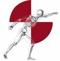
The Association of Osteomyologists foresaw this requirement for the advancement of experienced professional manual practitioners and sustains this progress through a training program of C.P.D. (Continuing Professional Development). Their logo shows the coming together of different bodies, meeting as one in the centre, and this is the whole concept of an Osteomyologist. He or she has decided that in order to treat their patients successfully, they have joined a group that believes in sharing each others expertise to the benefit of the patient.


Back Trouble UK


Tuesday 10 June 2008

Back Supports Are Not Always The Answer!

Spinal Lumbar or Lower Back Supports cannot really do anything to prevent pain in the lower back, a new review has reported.
The review, published in the latest issue of The Cochrane Library, found that the large belts which can be worn around the waist when picking up or carrying heavy objects are as effective in reducing pain as education about lifting.
The researchers looked at 15 studies which involved over 15,000 people and looked at both the prevention and treatment of low back pain. They found that patients who did not use such supports reported the same levels of pain or reduced disability as those who had.


Lead author Ingrid van Duijvenbode said that the findings of the review indicate that lumbar supports should not be recommended for patients looking to treat or manage their low back pain.
She said: "There is moderate evidence that lumbar supports do not prevent low back pain or sick leave more effectively than no intervention or education on lifting techniques in preventing long-term low back pain. There is conflicting evidence on the effectiveness of lumbar supports as treatment compared to no intervention or other interventions."

A spokesman for the Arthritis Research Campaign, which funds a great deal of research into alleviating low back pain, said that in most cases, patients benefited from maintaining everyday activities and keeping as mobile as possible, rather than resting.
Unfortunately back pain tends to deter exercise, but medical opinion is now firmly in favour of movement and exercise as part of back care.


Top Tips:
DO & DON'T
Select a form of exercise suitable for your fitness level - if you are a beginner, work up gradually. Don't do much in your first few days. Your tolerance will be very low at first, and if you do too much you won't be able to do anything the next day. Also, you will be put off.


Find an exercise that you enjoy. You will be more likely to continue. Don't do impact sports like running, at least to start with.


Learn to stretch. Stretches should be done slowly and gently, without jerks or excessive force. Don't just copy others, especially athletes! To get it really right, book a session with a physiotherapist. Don't do sports which make you bend your back, or move suddenly, like cycling or squash, until your back is fully recovered.


Stop if an activity creates increasing pain in your back. Find a different sport until you are fitter. Don't waste money on a home workout machine. Most people quickly stop using them because they are so boring.


Wear good quality trainers. These are excellent for reducing impact forces with the ground, which otherwise generate a shockwave up the back. Don't avoid lifting altogether. Moderate loads on your back are needed to build up fitness in the back muscles. But lift correctly.


Don't play competitive team games to start with - you may be tempted to overdo it, or you might hurt your back in a collision.


The best type and intensity of exercise is different according to your condition, your fitness and the state of your back. You need some exercise, but not too much. That's why you may be given conflicting advice by different experts. If that happens, learn the broad principles and then go your own way - it's your body and only you can feel it.


The key is to start gently, choose your sports carefully, and gradually develop into a more strenuous regime. Getting fit is a stress/recovery process, while most bad backs result from too much or too prolonged stress, with incomplete recovery. Your exercise "sweet spot" will be unique to you, and will change as you get fitter and your back heals.


Find something that you enjoy and that can be easily incorporated into your daily life. This can be easy to achieve, for instance: if you normally get the bus to work, try getting an earlier one and getting off a few stops earlier. Park your car further away from your destination and walk.


When going out to lunch, try to find a restaurant that is within walking distance. Walk up or down stairs instead of taking the lift.


Why exercise is good for your back:
When abdominal and back muscles are toned, they work together like a natural corset for your back, providing support and improving posture.
Fit muscles have better fine control and more power in reserve, giving smoother motion during lifting and moving.
Fitness postpones fatigue, helping you avoid tired habits like not lifting correctly.
Exercise makes bones denser and stronger.
With stretching, exercise increases flexibility, helps you use good postures for lifting and sitting.
Helps you lose weight and stop smoking! Excess body weight and smoking are bad for backs.
Exercise improves blood circulation, and so enhances nutrition and recovery processes in your back.

Terry O’Brien
BackTrouble.co.uk


Wednesday 21 May 2008

Homeopathy & Back Pain

Back pain ranks second only to headaches as the most frequent pain location.
Four out of five adults will experience at least one bout of back pain at some time in their life.
Back pain can occur for no apparent reason and at any point on your spine.

The most common site for pain is your lower back because it bears the most weight and stress.

Although back pain is common, it's also quite possible for you to prevent most back problems with simple steps such as exercise and adopting new ways to sit and stand.

Even if you've injured your back before, you can learn techniques to help avoid recurrent injuries.

Limited rest combined with appropriate exercise and education is often the best course.
Acute back pain often goes away by itself in a few days or weeks.
An ice bag or hot water bottle applied to the back may also help to alleviate pain. Prolonged bed rest is not beneficial because it weakens muscles.

Exercising with Back Pain
Recommendations for preventing initial and recurring episodes of back pain include:
regular exercise stretching before participation in sporting activities
losing weight maintaining correct posture
using comfortable, supportive seats while driving
sleeping on the side with knees drawn up or on the back with a pillow under bent knees

Homeopathy

Many of the herbs used for pain relief use the same biochemical pathways as the non-opiate pain-relieving drugs, but they are not as effective. However, on the positive side, many of these herbs have multiple effects. Their antispasmodic and circulation-promoting constituents may make up for what these plants lack in prostaglandin- suppressing strength. Herbal formulas that combine prostaglandin-suppressing, antispasmodic, sedative, and antidepressant plants are commonly prescribed by professional herbalists in North America, Great Britain, and Australia

Chronic pain often creates other problems besides the pain itself. These may include: tension, spasm, insomnia, and depression. And while conventional pain medications may remedy one or two of these side effects, some formulas of herbs can address them all. A pain-reliever, an antispasrnodic, a sedative, and an antidepressant may all be in included in a typical herbal formula created by a medical herbalist. For example, one herbal combination may include equal parts of willow bark (for pain), cramp bark (for spasm), valerian (a sedative), and St. Johns wort (an antidepressant).

For example, if related to drink. .

Hot, moist herbal packs help relieve the pain and increase blood circulation on painful areas, while herbal teas, juices and extracts soothe muscles and nerves.

Camomile has a calming effect on smooth muscle tissue. Take it as 1-3 cups of tea, 10-20 drops of extract in a cup of liquid or 1-3 capsules daily.

Bromelain (pineapple extract) is a powerful anti-inflammatory (take 2-3 g daily at first, then 1-2 g as the pain eases). Other anti-inflammatories, effective when drunk as teas, are valerian, St. John's wort, and Jamaican dogwood.

Horsetail not only heals and builds connective tissue, but also normalizes the bowels and alleviates lower-back pain, much of which can be traced to a dysfunctional intestinal tract. Take internally as per camomile.

Burdock soothes the pain and purifies the blood. Take 1-3 capsules or 10-25 drops of extract in 1 cup liquid daily.

If the muscle tension is due to emotional stress, take borage, St. John's wort, lemon balm or valerian teas.

Fresh yarrow juice is excellent for strengthening back muscles.

Use a white or black mustard seed pack for more intense heat. A mustard pack should not be left on for more than ten minutes because it can irritate the skin.

An infusion of meadowsweet three times a day combined with a rub on the area with lobelia and cramp bark is useful for physical strain or rheumatic problems.

Here are some herbs that are useful in pain relief.

Hot Peppers
Cayenne pepper (Capsicum spp.) is used in formulas for liniments and plasters in the folk medicine. Red pepper contains a pain-relieving chemical--capsaicin--that is so potent that a tiny amount provides the active ingredient in some powerful pharmaceutical topical analgesics. One product, Zostrix, contains only 0.025 percent capsaicin.

The exact mechanism in which red pepper works is not known. But it sure does work. Red pepper's effectiveness may be due to:

Capsaicin interferes with our pain perception
Capsaicin trigger release of the body's own pain-relieving endorphins
Salicylates present in red pepper.

How to Apply

1. You can buy a commercial cream containing capsaicin and use that.

2. Mash a red pepper and rub it directly on the painful area.

3. Take any white skin cream that you have on hand such as cold cream. Mix in enough red pepper to turn it pink.

4. Place 1 ounce of cayenne pepper in a quart of rubbing alcohol. Let the mixture stand for three weeks, shaking the bottle each day. Then, apply to the affected part during acute attacks.

5. Place 1 ounce of cayenne pepper in a pint of boiling water. Simmer for half an hour. Do not strain, but add a pint of rubbing alcohol. Let cool to room temperature. Apply as desired to the affected part.

Caution: Do not ingest any of these remedies. Wash your hands thoroughly after preparing or using red pepper. Don't get it in your eyes.

Some people are sensitive to this compound. Test it on a small area of skin to make sure that it's okay for you to use before using it on a larger area. If it seems to irritate your skin, discontinue use.

Cramp Bark and Black Haw
For the treatment of spasmodic pain, both cramp bark (Viburnum opulus) and black haw (Viburnum prunifolium) have been used in American Indian medicine. The Indians used cramp bark to treat both menstrual pain and muscle spasm. Cramp bark and black haw were also used hisatorically for arthritic or menstrual pain. The plants contain the antispasmodic and muscle-relaxing compounds esouletin and scopoletin. The antispasmodic constituents are best extracted with alcohol. So use tinctures rather than teas. Black haw also contains aspirin- like compounds.

Directions: Mix equal parts of cramp bark and black haw tinctures. Take between 1 and 4 droppers every two or three hours for up to three days.

Willow Bark
Willow bark (Salix alba) was used for treating pain by the ancient Greeks more than 2,400 years ago. American Indians throughout North America used it as a pain reliever even before the arrival of the European colonists. Investigation of salicin, a pain-relieving constituent in willow bark, led to the discovery of aspirin in 1899. The most important active constituent is salicin, but other anti-inflammatory constituents also appear in the willow bark.

Peppermint (Mentha piperita) and other mints.
The compounds menthol and camphor are found in many over-the-counter backache medications. They are chemicals that can help ease the muscle tightness that contributes to many bad backs. Menthol is a natural constituent of plants in the mint family, particularly peppermint and spearmint, although the aromatic oils of all the other mints contain it as well. Camphor occurs in spike lavender, hyssop and coriander.

Ginger
Ginger is used to treat various sorts of pain in the folk medicine of China and India. It is an important pain medication in contemporary Arabic medicine. Ginger contains 12 different aromatic anti-inflammatory compounds, including some with mild aspirin-like effects.

Directions: Cut a fresh ginger root (about the size of your thumb) into thin slices. Place the slices in a quart of water. Bring to a boil, and then simmer on the lowest possible heat for thirty minutes in a covered pot. Let cool for thirty more minutes. Strain and drink 1/2 to 1 cup, sweetened with honey, for taste if needed.

Rosemary
Drinking rosemary tea for pain is a remedy used in the contemporary Hispanic folk medicine of Mexico and the Southwest. Its leaf also contains four anti-inflammatory substances---camosol, oleanolic acid, rosmarinic acid, and ursolic acid. Carnosol acts on the same anti-inflammatory pathways as both steroids and aspirin; rosmarinic acid acts through at least two separate anti-inflammatory biochemical pathways; and ursolic acid, which makes up about 4 percent of the plant by weight, has been shown in animal trials to have anti-arthritic effects.

Directions: Put 1/2 ounce of rosemary leaves in a 1-quart canning jar and fill the jar with boiling water. Cover tightly and let it stand for thirty minutes. Drink a cup as hot as possible before going to bed, and have another cupful in the morning before breakfast.

Epsom Salt Baths
Folk traditions call for Epsom salt baths to relieve pain. Epsom salt was reputed to have magical healing properties. Epsom salt is primarily magnesium sulfate and has been used medicinally in Europe for more than three hundred years. The heat of an Epsom salt bath can increase circulation and reduce the swelling of arthritis, and the magnesium can be absorbed through the skin. Magnesium is one of the most important minerals in the body, participating in at least 300 enzyme systems. Magnesium has both anti-inflammatory and anti-arthritic properties.

Directions: Fill a bathtub with water as hot as can be tolerated. Add 2 cups of Epsom salts. Bathe for thirty minutes, adding hot water if necessary to keep the bath water warm.

Angelica
Various species of angelica have been used to quiet pain by American Indians throughout North America. The European species (Angelica archangelica) and the Chinese species (Angelica sinensis) have been used in the same way in the folk medicine of Europe and China respectively. The Chinese species is sometimes sold in North America under the names dang gui or dong quai. All species contain anti-inflammatory, antispasmodic, and anodyne (pain- relieving) properties. The European species of angelica has been used in European folk medicine since antiquity, as has the Chinese species in Chinese medicine.

Directions: Place 1 tablespoon of the cut roots of either species of angelica in a pint of water and bring to a boil for two minutes in a covered pot. Remove from heat and let stand, covered, until the tea cools to room temperature. Drink the pint in 3 doses during the day.

What ever you decide to do with regard to treating your medical condition, always consult with your GP before starting any alternative therapies or medicines.

Thursday 8 May 2008

Coping with a Whiplash Neck Injury.

Even with the many advances in medicine and in particular skeletomuscular medicine, Whiplash is still not that well understood. Severity of injury varies considerably between people in the same types of car accidents, even when circumstances are similar.

Speed is a major factor, as is the weight of the vehicles.

There are several other factors involved. When the person sees the accident coming, the injury is often less severe. This may be because the nervous system has time to prepare.
The position of the neck at the time of impact also plays a role. Risk of injury appears to be greater when the head is turned to the side than when it is facing straight ahead - except in very low speed crashes. The position of the headrest is important - it needs to be at the proper height and distance from the head to reduce severity of injury. The strength of a person’s neck also plays a role. Females, having less strength in their necks, generally suffer more severe whiplash injuries than males.
Seatbelts with shoulder harnesses should always be used and head rests at the proper height for the person. The height of the headrest should be just above the ear. The distance between the headrest and back of the head should be two to four inches.
Symptoms may occur immediately after the injury or set in gradually over the next couple of days. It is common for a person with no immediate symptoms to wake up stiff and sore the next morning. Whiplash can vary greatly in severity and cause a wide range of symptoms. Not all people experience the same symptoms. Symptoms that develop rapidly often indicate a serious injury.

The most common symptoms of whiplash are neck pain and stiffness. The neck becomes stiff as muscles tighten up to protect the injured area from further injury by reducing motion.
Headaches are also common. Pain originating in the neck (from muscle spasms in the neck and/or irritated nerves in the back of the neck) is often referred to the head. The pain may felt be over the entire head or any area of the head - often over the forehead and behind the eyes.
There may be back pain or shoulder pain, or numbness or tingling in the arm. Other symptoms of whiplash include problems with memory and concentration, feelings of disorientation, dizziness, ringing in the ears, impaired hearing, blurred vision, sensitivity to sound and light, irritability, depression, and difficulty sleeping.

Neurological symptoms may be the result of injury to soft tissue injury to the neck or a mild brain injury or concussion. A sudden jolt to the head can jar the brain. Neurological symptoms often resolve within a week.
If symptoms occur immediately or shortly after the accident an ambulance should be called so that the neck can be immobilised during transport to a hospital emergency ward.
There may be serious injuries that may require immediate medical treatment (such as haemorrhage, fracture, dislocation, or spinal cord injury). In addition, auto accidents that result in whiplash may also result in other injuries such as chest injuries, back injuries, internal injuries, etc.

Always seek a proper diagnosis from a GP, even if the symptoms are mild. The diagnosis may be based upon symptoms alone, or an MRI or CT may be taken to see the extent of soft tissue injury. X-rays may be taken to rule out fractures or dislocation.

Treatment depends on severity of symptoms. If the injury is mild, applying ice packs at home for the first couple of days along with the short-term use of NSAIDs may be sufficient. If symptoms are moderate (especially if there is limited range of motion), physical therapy is often recommended. Massage therapy and/or spinal manipulation may also be beneficial.
Applying IceApplying ice every four hours for the first couple of days helps reduce inflammation (the main cause of the pain). Inflammation is greatest for the first two days. Wrap ice in a cloth and apply to area of neck pain for 20 minutes every three to four hours.

Medications
Anti-inflammatory medication to relieve both pain and inflammation such as Ibuprofen or Aspirin are often helpful. Your GP may prescribe muscle relaxants or pain medication that contains codeine for temporary use.

Physical Therapy
Depending upon the severity of the whiplash injury, physical therapy may be recommended by your GP. A physical therapist can prescribe range-of-motion exercises, teach proper posture to avoid excess strain on neck, treat pain with cold and heat, etc.
Prolonged Use of Soft Collars do Delay Recovery.
Soft collars may be helpful for the first two or three days, when pain and inflammation are at its greatest. Inflammation may trigger muscle spasms - a protective mechanism to restrict movement to prevent further injury. Wearing a soft collar helps relax the muscles to relieve pain. However, prolonged use of soft collars has been shown to delay recovery from whiplash. Intermittent use may be recommended in some cases.
*If a fracture or dislocation is involved, a wearing moulded collar may be necessary to stabilize the neck.

How quickly one recovers from whiplash depends largely upon the severity of the injury. How quickly the symptoms develop often correlate with the seriousness of the injury.
Mild whiplash injuries often heal completely within two to three weeks, moderate whiplash injuries within two to three months. Severe whiplash injury may take several months to heal. For some people, symptoms (such as mild neck pain and/or headaches) may linger for longer periods of time. If symptoms are still present after six months, the pain is considered to be chronic. Chronic symptoms may last for years. Whiplash injury may increase risk of degenerative changes in the discs and spinal joints.


Their have been cases were people report that new symptoms appear years after the initial whiplash injury, though it is difficult to determine whether or not symptoms that appear years later are related to the whiplash injury.

Terry O’Brien
BackTrouble.co.uk


Monday 28 April 2008

Degenerative Joint Disease.

Alternative Names: Hypertrophic osteoarthritis; Osteoarthrosis; Degenerative joint disease; DJD; OA; Arthritis-Osteoarthritis.

Causes
Unfortunately most of the time, the cause of OA is unknown. It is mainly related to aging, but metabolic, genetic, chemical, and mechanical factors can also lead to OA.
The symptoms of osteoarthritis usually appear in middle age and almost everyone has them by age 70. Before age 55, the condition occurs equally in both sexes. However, after 55 it is more common in women.

The disease causes the cushioning (cartilage) between the bone joints to wear away, leading to pain and stiffness. As the disease gets worse, the cartilage disappears and the bone rubs on bone. Bony spurs usually form around the joint.

OA can be primary or secondary.
Primary OA occurs without any type of injury or obvious cause.
Secondary OA is osteoarthritis due to another disease or condition. The most common causes of secondary OA are metabolic conditions, such as acromegaly, problems with anatomy (for example, being bow-legged), injury, or inflammatory disorders such as septic arthritis.

Symptoms
The symptoms of osteoarthritis include:
Deep aching joint pain that gets worse after exercise or putting weight on it and is relieved by rest
Grating of the joint with motion
Joint pain in rainy weather
Joint swelling
Limited movement
Morning stiffness
Of course some people might not have symptoms.
Examination and Tests
A physical exam can show limited range of motion, grating of a joint with motion, joint swelling, and tenderness.
An x-ray of affected joints will show loss of the joint space, and in advanced cases, wearing down of the ends of the bone and bone spurs.

Treatment
The goals of treatment are to relieve pain, maintain or improve joint movement, increase the strength of the joints, and reduce the disabling affects of the disease. The treatment depends on which joints are involved.

MEDICATIONS
The most common medications used to treat osteoarthritis are nonsteroidal anti-inflammatory drugs (NSAIDs). They are pain relievers that reduce pain and swelling. Types include aspirin, ibuprofen, and naproxen.
Although NSAIDs work well, long-term use of these drugs can cause stomach problems, such as ulcers and bleeding. Manufacturers of NSAIDs include a warning label on their products that alerts users to an increased risk for cardiovascular events (heart attacks and strokes) and gastrointestinal bleeding.

Other medications used to treat OA include:
COX-2 inhibitors (coxibs). Coxibs block a substance called COX-2 that causes swelling. This class of drugs was first thought to work as well as other NSAIDs, but with fewer stomach problems. However, reports of heart attacks and stroke have led the Drug Licensing Authorities to re-evaluate the risks and benefits of the COX-2s. Ask your doctor whether the drug is 1. Still available and if so 2. Right and safe for you.
Steroids. These medications are injected right into the joint. They can also be used to reduce inflammation and pain.

Supplements. Many people are helped by over-the-counter remedies such as glucosamine and chondroitin sulfate. There is some evidence that these supplements can help control pain, although they do not seem to grow new cartilage.
Artificial joint fluid (Synvisc, Hyalgan). These medications can be injected into the knee. They may relieve pain for up to 6 months.

LIFESTYLE CHANGES
Exercise helps maintain joint and overall movement. Ask your health care provider to recommend an appropriate home exercise routine. Water exercises, such as swimming, are especially helpful.
Applying heat and cold, protecting the joints, using self-help devices, and rest are all recommended.
Good nutrition and careful weight control are also important. If you're overweight, losing weight will reduce the strain on the knee and ankle joints.

PHYSICAL THERAPY
Physical therapy can help improve muscle strength and the motion at stiff joints. Therapists have many techniques for treating osteoarthritis. If therapy does not make you feel better after 3-6 weeks, then it likely will not work at all.

BRACES
Splints and braces can sometimes support weakened joints. Some prevent the joint from moving; others allow some movement. You should use a brace only when your doctor or therapist recommends one. Using a brace the wrong way can cause joint damage, stiffness, and pain.

SURGERY
Severe cases of osteoarthritis might need surgery to replace or repair damaged joints. Surgical options include:
Total or partial replacement of the damaged joint with an artificial joint (knee arthroplasty, hip arthroplasty )
Arthroscopic surgery to trim torn and damaged cartilage and wash out the joint
Cartilage restoration to replace the damaged or missing cartilage in some younger patents with arthritis
Change in the alignment of a bone to relieve stress on the bone or joint (osteotomy)
Surgical fusion of bones, usually in the spine (arthodesis)

Outlook (Prognosis)
Your movement may become very limited. However Treatment generally improves function.
Possible Complications
Decreased ability to walk
Decreased ability to perform everyday activities, such as personal hygiene, household chores, or cooking
Adverse reactions to drugs used for treatment
Surgical complications
When to Contact a Medical Professional
Do contact your health care provider if you have symptoms of osteoarthritis.

Prevention
*Weight loss can certainly help in the management of DJD and reduce the risk of knee osteoarthritis in overweight women.

Degenerative Joint Disease

Alternative Names: Hypertrophic osteoarthritis; Osteoarthrosis; Degenerative joint disease; DJD; OA; Arthritis-Osteoarthritis.
Causes
Unfortunately most of the time, the cause of OA is unknown. It is mainly related to aging, but metabolic, genetic, chemical, and mechanical factors can also lead to OA.
The symptoms of osteoarthritis usually appear in middle age and almost everyone has them by age 70. Before age 55, the condition occurs equally in both sexes. However, after 55 it is more common in women.


The disease causes the cushioning (cartilage) between the bone joints to wear away, leading to pain and stiffness. As the disease gets worse, the cartilage disappears and the bone rubs on bone. Bony spurs usually form around the joint.


OA can be primary or secondary.
Primary OA occurs without any type of injury or obvious cause.
Secondary OA is osteoarthritis due to another disease or condition. The most common causes of secondary OA are metabolic conditions, such as acromegaly, problems with anatomy (for example, being bow-legged), injury, or inflammatory disorders such as septic arthritis.
Symptoms
The symptoms of osteoarthritis include:
Deep aching joint pain that gets worse after exercise or putting weight on it and is relieved by rest.
Grating of the joint with motion.
Joint pain in rainy weather.
Joint swelling.
Limited movement.
Morning stiffness.
Of course some people might not have symptoms.

Examination and Tests
A physical exam can show limited range of motion, grating of a joint with motion, joint swelling, and tenderness.
An x-ray of affected joints will show loss of the joint space, and in advanced cases, wearing down of the ends of the bone and bone spurs.

Treatment
The goals of treatment are to relieve pain, maintain or improve joint movement, increase the strength of the joints, and reduce the disabling affects of the disease. The treatment depends on which joints are involved.

MEDICATIONS
The most common medications used to treat osteoarthritis are nonsteroidal anti-inflammatory drugs (NSAIDs). They are pain relievers that reduce pain and swelling. Types include aspirin, ibuprofen, and naproxen.

Although NSAIDs work well, long-term use of these drugs can cause stomach problems, such as ulcers and bleeding. Manufacturers of NSAIDs include a warning label on their products that alerts users to an increased risk for cardiovascular events (heart attacks and strokes) and gastrointestinal bleeding.
Other medications used to treat OA include:
COX-2 inhibitors (coxibs). Coxibs block a substance called COX-2 that causes swelling. This class of drugs was first thought to work as well as other NSAIDs, but with fewer stomach problems. However, reports of heart attacks and stroke have led the Drug Licensing Authorities to re-evaluate the risks and benefits of the COX-2s. Ask your doctor whether the drug is 1. Still available and if so 2. Right and safe for you.


Steroids. These medications are injected right into the joint. They can also be used to reduce inflammation and pain.
Supplements. Many people are helped by over-the-counter remedies such as glucosamine and chondroitin sulfate. There is some evidence that these supplements can help control pain, although they do not seem to grow new cartilage.
Artificial joint fluid (Synvisc, Hyalgan). These medications can be injected into the knee. They may relieve pain for up to 6 months.


LIFESTYLE CHANGES
Exercise helps maintain joint and overall movement. Ask your health care provider to recommend an appropriate home exercise routine. Water exercises, such as swimming, are especially helpful.
Applying heat and cold, protecting the joints, using self-help devices, and rest are all recommended.
Good nutrition and careful weight control are also important. If you're overweight, losing weight will reduce the strain on the knee and ankle joints.


PHYSICAL THERAPY
Physical therapy can help improve muscle strength and the motion at stiff joints. Therapists have many techniques for treating osteoarthritis. If therapy does not make you feel better after 3-6 weeks, then it likely will not work at all.

BRACES
Splints and braces can sometimes support weakened joints. Some prevent the joint from moving; others allow some movement. You should use a brace only when your doctor or therapist recommends one. Using a brace the wrong way can cause joint damage, stiffness, and pain.
SURGERY
Severe cases of osteoarthritis might need surgery to replace or repair damaged joints. Surgical options include:
Total or partial replacement of the damaged joint with an artificial joint (knee arthroplasty, hip arthroplasty )
Arthroscopic surgery to trim torn and damaged cartilage and wash out the joint
Cartilage restoration to replace the damaged or missing cartilage in some younger patents with arthritis
Change in the alignment of a bone to relieve stress on the bone or joint (osteotomy)
Surgical fusion of bones, usually in the spine (arthodesis)

Outlook (Prognosis)
Your movement may become very limited. However Treatment generally improves function.
Possible Complications.
Decreased ability to walk.
Decreased ability to perform everyday activities, such as personal hygiene, household chores, or cooking.
Adverse reactions to drugs used for treatment.
Surgical complications.

When to Contact a Medical Professional
Do contact your health care provider if you have symptoms of osteoarthritis.

Prevention
*Weight loss can considerably help in the management DJD and ceratainly reduce the risk of knee osteoarthritis in overweight women.


Friday 18 April 2008

Bio-Mechanics and Back Pain Relief.

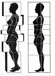
When our body is in the standing position it is in a state of unsteady equilibrium because the base is small and the centre of gravity is a long way above it. That is why it has to be balanced by keeping the centre of gravity over the base, otherwise the body would fall over.Equilibrium: The steadiness of the body depends on the size of its base and the distance of the centre of gravity from that base. An object having a large base and a low centre of gravity is not easily upset.The first thing necessary in order to accomplish good posture is to straighten the neck, keep the chin down and back.

The second important thing is correct deep breathing. The line of gravity in the fundamental standing position falls in front of the ears, and in front of the cervical and dorsal vertebrae, through the lumbar vertebrae, through the hip joint, and slightly in front of the ankle joint. But this probably varies in individuals according to their build and height."Biomechanics is that branch of science concerned with the understanding of the interrelationships of structure and function of living beings with respect to the kinematics and kinetics of motion"Poor Posture associated by the following:

Genetic disorders, Familial association, Irregular/no exercise, Bad habits when sitting or standing, Carrying excess loads, (School/shopping) Underweight/Overweight (Inadequate diet)Mental Attitude towards oneself.

Biomechanics is our way of describing how each bit of your body moves in relation to another bit.
Our bodies are fantastic at compensating when things are not quite perfect but unfortunately when we run out of compensations we usually end up with pain and stiffness.


Let’s have a look at some of the more common biomechanical problems:

Problem 1.
It’s the fashion not to tie trainers and shoes up but this prevents the shoe from supporting the inside of the foot and allows the foot to roll or ‘pronate’ too much in standing and walking.
This means the knee and the hip rolls inwards too much which makes the inside calf and inside thigh muscle shorter. The iliotibial band can then rub against the outside knee giving pain.
Further up, the bottom muscles become too long and stop working as they should do which puts added stress on the pelvis and low back joints. Before you know it you have back pain, knee pain and ankle pain! And all because of fashion!

Problem 2.
Whether you drive 5 miles or 500 miles a day your car seat is really important. In many cars the base of the seat slopes backwards. In fact some of the more expensive cars are the worst because they want you to feel ‘snug’ and safe in the seat. However, this position forces your pelvis to sit too far backwards.


Once this happens your low back and upper back flex forward too much but then your neck muscles have to work really hard to stop your head from flexing too much and allow you to see where you are going. This position then forces your shoulder blades to stretch too far forwards and a large stress is placed on both the front and the back of the shoulder.
The result is neck pain and /or upper or low back pain. This position can exacerbate sciatica as it is a big stretch for the nerves.


The solution is to raise the back of the seat so it becomes more horizontal. Some cars allow you to alter this angle but if yours does not then sit on a small cushion or buy a special wedge shape cushion that fits into the seat.

Terry O’Brien
Back Trouble UK.

Thursday 10 April 2008

The Silent Disease


The Silent Disease is the name that is often given to Osteoporosis, as most people don't know they have osteoporosis until it has progressed often to the point of fracture, usually in the hip, wrist or spine. Even when undiagnosed osteoporosis results in a vertebral fracture, the pain is frequently dismissed as general back pain. This lack of awareness can lead to serious illness, deformity, even death.

Osteoporosis alone does not cause back pain. It can, however, weaken the spine to where it is no longer able to withstand normal stress or minor trauma, resulting in vertebral fracture. It is the ensuing fracture that causes pain
.

Osteopenia is a condition where bone mineral density is lower than normal, but not low enough to be classified as osteoporosis. While often a precursor to osteoporosis, not everyone with osteopenia will develop osteoporosis. Since a diagnosis of osteopenia puts one at greater risk for osteoporosis, patients are encouraged to seek the advice of their physicians about implementing preventive measures.

Osteoporosis Causes and Risk Factors
Bones are made of complex, constantly changing, living tissue. They are able to grow and heal, and are also susceptible to changes in diet, body chemistry, and exercise levels.
Early in life, more bone is laid down than is removed by the body. People typically achieve peak bone mass by around age 30, after which more bone is lost than is replaced. Too much bone loss leads to osteoporosis.

Both of the two primary types of osteoporosis are far more common in women than men:

Type I osteoporosis (postmenopausal osteoporosis) —generally develops after menopause, when estrogen levels drop precipitously, leading to bone loss — usually in the trabecular (spongy) bone inside the hard cortical bone.


Type II osteoporosis (senile osteoporosis) — typically happens after age 70 and involves a thinning of both the trabecular (spongy) and cortical (hard) bone.

In addition, certain medications and medical conditions can damage bone and lead to what is known as “secondary osteoporosis”. Patients being treated for any of the following conditions should discuss the risk of osteoporosis with their physicians:

Endocrine disorders

Marrow disorders

Collagen disorders

Gastrointestinal disorders

Seizure disorders

Eating disorders (such as anorexia or bulimia)

It is important to distinguish between primary and secondary causes of osteoporosis because treatment is often different. To determine the cause, a thorough medical history, physical examination, and appropriate diagnostic tests need to be conducted (see Diagnosing Osteoporosis).

Key risk factors for developing osteoporosis include:

Advanced = age over age 65.

Gender = Women are four times more likely to develop osteoporosis than men.

Heredity = Family history of osteoporosis or fracture on the mother’s side.

Personal history = any type of fracture after age 45.

Race = Caucasian and Asian women are at greater risk.

Body type= small-boned women weighing less than 127 pounds.

Menstrual history = Normal menopause increases the risk of osteoporosis and early menopause can exacerbate this risk.

Lifestyle = calcium and/or vitamin D deficiency; little or no exercise (especially weight-bearing exercise); alcohol abuse; smoking; too much cola/soda.

Testosterone deficiency (hypgonadism) = in men.

Why women are at greater risk for developing osteoporosis

Estrogen plays an important part in maintaining bone strength. Starting at about age 30 through onset of menopause, women lose a small amount of bone every year as a natural part of the aging process. When women reach menopause and estrogen levels decrease, the rate of bone loss increases for approximately 8 to 10 years before returning to premenopausal rates.

Osteoporosis Symptoms

Osteoporosis can go undetected for years and fracture is typically the first outward sign. Advanced osteoporosis is potentially disabling, often leading to one or more of the following:
fractures of the spine, wrist or hip

spinal deformity (e.g., lost height, hunched back)

chronic or severe pain

limited function and reduced mobility

loss of independence

decreased lung capacity

difficulty sleeping

Osteoporosis is the leading cause of spine fractures, especially in women over age 50, but only about one third of all spine fractures are diagnosed.

Most osteoporotic spine fractures (vertebral compression fractures) start with sudden back pain, usually after routine activity (lifting or bending) that slightly strains or jars the back. After a month or two, this acute pain is usually replaced by an achy pain (see Diagnosing vertebral compression fractures).

Osteoporosis Prevention

Postmenopausal (Type I) osteoporosis can be significantly influenced by preventive measures. Most of these behaviors are up to the individual and should be started as early in life as possible. For those genetically predisposed to osteoporosis, the following practices are even more important:
Exercise regularly weight-bearing exercises (activities that work one’s bones and muscles against gravity) are essential to maintaining bone health.

Ensure adequate calcium intake, Calcium plays a key role in keeping bones strong. Vitamin D is also essential, as it helps ensure absorption and retention of calcium in bones. Calcium and vitamin D requirements vary depending on age and gender.

Eat a balanced, healthy diet Certain foods provide excellent sources of calcium, while diets high in protein and/or sodium increase calcium loss.

Quit smoking - Smoking has a detrimental effect on bone density, leading to greater risk of injury and longer recovery times.

Limit alcohol consumption - While the exact way alcohol affects bone isn’t entirely understood, excessive alcohol use has been proven to accelerate bone loss.

Limit intake of colas/sodas - recent research indicates that too much cola or soda can increase the risk of osteoporosis.

Undergo bone density testing - every 1-2 years if you are postmenopausal, over age 65, or have other risk factors. Bone mineral density (BMD) tests indicate normal, low or osteoporotic bone density levels, as well as any increased risk of fracture.

For more information, see How to prevent osteoporosis.

Osteoporosis Treatment

Once osteoporosis has been diagnosed, patient and physician should work together to develop a treatment plan where the goal is to slow bone loss and prevent fractures. Treatment may include:
Education on diet/nutrition - see Food for Thought: Diet and Nutrition for a Healthy Back.

Exercise (if no fracture) - to help maintain bone density and reduce the risk of falls.

Medication - to slow bone loss and prevent fractures. Osteoporosis medications fall into two categories:

medications that slow or stop bone resorption (loss);

medications that increase bone formation.

Treatment for vertebral fractures, which may include:

rest, though long-term rest accelerates bone loss;

rigid back braces to support the spine;

ice/heat and pain medications;

surgery (kyphoplasty or vertebroplasty), which may be necessary in certain situations where the fracture is causing severe pain and/or deformity, or has failed to respond to three months of non-surgical treatment.

Do be positive because even once osteoporosis has been diagnosed, it is possible to slow bone loss, build bone density and prevent fractures. Continually advancing osteoporosis and related fractures are not an inevitable outcome of being diagnosed with osteoporosis.


Terry O’Brien
Back Trouble UK.

Tuesday 25 March 2008

Arthritis

Spinal arthritis is a very painful condition, worse than other forms of arthritis due to the high amount of nerves present in and around the spinal structures, especially those concerning the facet joints and intervertebral discs.
Spinal osteoarthritis, otherwise known as spondylosis, can affect any region of the spine and is caused by the degenerative process.

More common in the over 55s and with women more than men, spinal arthritis can be triggered by an accident, injury or from a weakened immune system.
People who have a family history of spinal arthritis are more likely to develop the condition themselves.

Symptoms
Apart from suffering with high levels of pain, there is the likelihood of having an increased stiffness, especially in the spine, particularly after long periods of rest. Pain can be intermittent and worse on movement.
As part of the human body’s natural response to arthritis, an excess of bony growth may occur, resulting in ‘spurs’ being formed। They can cause additional pain and can restrict movement further. Some sufferers report a crunching sound, like bone on bone, possibly due to the growth of the spurs.

Spinal arthritis occurring in the lower back can affect the lower extremities with additional pain in the legs and buttocks. Cervical spine arthritis can cause pain to radiate to the shoulders and arms.
There is no known cure for arthritis but there are a number of treatments available for the relief of symptoms.

Treatment
The treating GP may refer you to an x-ray department in order to confirm diagnosis by a series of images। These pictures will also show any inflammation and if there is any deterioration in the cartilage.

Along with this a blood sample can be gained and sent for analysis to determine the type of arthritis. Medications include anti-inflammatory drugs, pain relief, arthritic drugs or steroids.
Spinal spondylosis can cause the intervertebral discs to degenerate which increases the risk of them herniating, a condition that would require further treatment.

Facet joints of the spine can be affected by the deterioration of cartilage or the formation of bony overgrowths and can limit movement significantly. Surgery may be an option to remove the bony growths.
Treatments that can be performed by the patient, without input from a medical professional include giving-up smoking; this will increase the levels of oxygen to the affected area and prevent further deterioration of the discs. Maintaining a healthy weight for your frame will prevent putting excessive pressure and strain on the ligaments and joints of the spine. Applying regular heat or ice therapy, whichever is preferred, will help with pain relief and reduce inflammation. Exercise is a good way of help alleviate symptoms, as arthritis can affect mobility, it may be best to seek advice from a physiotherapist who will work with you and devise an appropriate and manageable programme of exercise.

Spinal arthritis is a painful and restrictive condition. Though there is no cure, it may be prevented or onset delayed by eating a healthy diet, rich in nutrients and calcium (to keep bones strong and healthy), and by ensuring flexibility, strength and suppleness by keeping to a regular exercise regime.

Thursday 6 March 2008

Back Exercises

Many times individuals experience back pain because their body is not strong enough to support their bodies. The result of a weak body is often back pain. However, back pain exercises are a great way to strengthen the entire body and provide support for your back. When your back has support because your body is strong due to back pain exercises then you will not experience back pain like you did before. So, if you suffer from back pain consider back pain exercises to increase your back strength and decrease your pain.

Back Pain Exercises


Back pain exercises might sound as if they are exercises that only focus on the back; however this is not the case. In fact, back pain exercises are made up of exercises that strengthen the back, stomach, neck, hips and thighs. The reason back pain exercises focus on all of these muscles is because all of these muscles work together to provide support for your back. When all of these muscles are strong due to back pain exercises they will provide a sound support system for your spine and your back pain days will be over.


Back pain exercises should include cardiovascular exercises like biking, running, or swimming followed up by wall sits, lounges, leg raises, sit ups, and other similar back pain exercises. When these back pain exercises are performed on a regular basis the individual will gradually strengthen their body providing more support to their back and body. As the body becomes stronger and better supported back pain will subside.

Sunday 17 February 2008

The Alexander Technique


The Alexander Technique is used to help to teach people about how efficiently and effortlessly they can use their bodies in everyday life. Often, we develop bad posture and habits without being aware of this, and expend too much energy or muscle force to achieve a task. Alexander Technique teachers help to adjust the client's posture to recognise the difference between current habits and what it feels like to use muscles with minimum effort and in a relaxed, fluid way.


The Technique teaches how to become more aware of your own posture, balance and movement in everyday life. The lessons usually last for between 30 and 45 minutes and are normally on a one-to-one basis. The teacher uses his/her hands to gently correct any muscular imbalances and encourages the body to a better alignment. This is a direct body experience, so the client becomes familiar with the sensation of correct alignment in his or her own body. This can feel strange initially, as the body is not used to using it's muscles in this way, and the new methods of movement need to be practised with constant awareness as to how we choose to use our bodies in everyday tasks. It is a process of re-educating the body by learning how to stand and move correctly. This leads to health benefits as often-poor spinal posture will lead to other symptoms like poor breathing due to restriction in the throat and diaphragm areas. Breathing and how we breathe is an important aspect of the Alexander Technique.


An Australian actor, Frederick Matthias Alexander (1869-1955), developed the Alexander Technique. He suffered from respiratory problems as a child, which later affected his voice and career in the theatre. He tried many remedies without success and eventually began a process of self-observation to try to find a way of curing himself. He realised that the voice problem was a result of muscular tension in his whole body and that his thought patterns also had a great part in contributing to the tension that had become an ingrained habit. Alexander studied his posture with the aid of mirrors to see how this was affected when he recited and as a result could see that his body alignment was incorrect. He gradually taught himself to correct his posture and found that he had cured his voice problem. He went on to pass the technique to others and eventually opened a clinic to help people to learn about their own use of posture.

Tuesday 12 February 2008

Back Pain Treatment without Drugs!


C’mon there is a standard "cure" for most causes of back and neck pain?


Fact: Compared to other medical conditions, there are relatively few standardised approaches to diagnosis and treatment of back problems. Spine specialists from various areas of expertise (such as physical medicine and rehabilitation, chiropractic, osteopathic medicine, physical therapy and surgery) will often disagree on the diagnosis and most appropriate treatment plan for back pain and back problems, and specialists within a discipline will also frequently have different opinions.


A few diagnoses for back pain as a result of back a back problem are relatively straightforward (such as a spinal tumour, infection, or fracture) and there is generally more consensus for diagnosis and treatment of these back problems.


Myth:
Rest is the key to recovery from back pain and back problems
Fact: The two main reasons bed rest may be recommended for back pain are to reduce pressure on the discs in the spine and to stop the mechanical stresses that are irritating pain receptors. A short period of bed rest may help reduce acute back pain. However, in most instances, more than 1 or 2 days of rest can be detrimental to recovery from back pain, potentially leading to increased pain and other adverse results, such as:


muscle atrophy (1% to 1.5% per day)


cardiopulmonary reconditioning (15% loss in 10 days)


bone mineral loss


risk of blood clots


loss of wages


creating an "illness" mindset


Myth:


Heat and massage feel good so they must be helping back pain and back problems?


Fact: These therapies can reduce acute back pain in the short-term, but do not provide a long-term solution to back problems. They are used to manage pain during recovery, allowing patients to complete a rehabilitation program and participate in daily activities.


Myth:


Long-term pain indicates I need back surgery for my back problems?


Fact: If back pain has reached the chronic stage, spine surgery actually has a reduced likelihood of being successful. Typically, symptoms that suggest back surgery might be helpful occur early in the course of pain and are relatively apparent.

Thursday 24 January 2008

Let us Help?

Are you a candidate for treatment?

Our associated clinics treat both acute and chronic back conditions.

If your symptoms have appeared recently we can use the best manual physical

therapy techniques to facilitate the healing process and restore normal function as soon as possible.

If your symptoms are long-standing (more than 6 weeks) or recurrent you are likely to have developed weakness and instability in your spine and you may be a candidate for our specialised programme of intensive strengthening of the lumbar spine.

We are able to treat anyone between the ages of 18 to 80.
Treatment is as suitable for those with sedentary lives as it is for athletes - we have patients at both ends of the scale.

This list shows some of the the conditions that can be treated successfully using Natural Physical Therapy.

Lumbar strain

Herniated (prolapsed) disc

Sciatica

Degenerative disc (or joint) disease

Stenosis

Spondylolisthesis

Osteoporosis

Osteoarthritis

Hypermobility/instability

Scheuermann's disease

Many treatment methods help only with symptomatic relief but do not address the underlying cause of pain and therefore result in only a temporary solution.

That is why so many people with back pain find themselves in the "Pain Maze" confused over which therapy to undergo, or whether drugs, injections, manipulation or surgical procedures will indeed bring an end to the pain.

That’s why Back Trouble UK was formed, so that Back Pain sufferers could be both more informed and to be able to make a more informed choice of available therapies.

Friday 11 January 2008

Craniosacral Therapy Whats it Mean?


Let me introduce Craniosacral Therapy, it is a non-invasive form of treatment that has its roots in the earliest days of Osteopathy, but has since grown and developed in its own right to become a powerful, yet subtle form of treatment.

At the beginning of the twentieth century, Dr. William Garner Sutherland, the founder of Cranial work, discovered that the different fluids and tissues (e.g. bones, muscles, organs etc.) of the body express a natural rhythmic motion that can be perceived through a light "listening" touch and can give the practitioner a wealth of information about the functioning of the body. This natural motion is considered to be a direct expression of the state of health of the individual.

Craniosacral therapy works through the practitioner helping to directly influence this most fundamental expression of health within the client. As such, it can be of great benefit to help relieve many physical, psychological and emotional conditions. Unlike other therapies, Craniosacral therapists do not try to directly resolve any symptomatic issues that might be present. Rather, they try to increase the level of available health within the affected area, and the body as a whole by re-establishing natural motion to the problem area. Due to the different experiences of our lives, these natural rhythmic motions may become restricted, or otherwise disturbed.

The more intense the experience (e.g. accidents, i llnesses, surgery, stress, trauma etc.), the greater the effect upon our body. This may lead to the development of various symptoms including reduced blood flow, increased levels of stress, lowered immunity, increased body tension as well as increased pain and dysfunction. It has been found that the most effective way to influence these natural motions, and to help restore the body to its optimum health, is through a light, non-invasive touch, without the use of massage, manipulation or other such techniques.

Where areas of restriction are found, the therapist will subtly encourage or enable the body's own potent healing forces to re-establish themselves in those places, leading to an overall return of healthy movement and function.

Wednesday 2 January 2008

First Aid for Back Pain!


Apply Heat and Ice:

Apply an ice pack to the affected area. If you haven't got an ice pack, use a bag of frozen vegetables instead. Don't put the ice directly on your skin, as it might cause a cold burn. Put a wet cloth between the ice and your skin.

If ice doesn't work, try applying gentle warmth with a hot water bottle. Don't apply the hot water bottle directly to the skin - cover it so it isn't too hot. A hot bath or shower might also help.

Some people find alternating heat and cold produces most relief. Try to get professional advice on applying heat and ice if you can.

Medication:

Take painkillers following the instructions on the packet - never take more than the recommended dose. Many people find that paracetamol or ibuprofen helps - your pharmacist can advise you.

Painkillers shouldn't be used as a long-term solution. If you find you still need them after a week or so, consult your doctor.

Relaxation:

Muscle tension is bad for back pain, so try to relax as much as possible. Take a long bath or listen to soothing music. Use a relaxation tape if you have one.

A gentle massage from a partner or friend may help, but make sure they don't do anything that causes pain.

Bed rest versus exercise:

Doctors used to recommend long periods of rest for people with backache, but research has shown this is actually bad for backs. Even crawling around on your hands and knees is better than no movement at all.

Some kinds of exercise, such as walking, don't put too much stress on your back. It's a good idea to make a start on them even if your back is a bit sore, just to get your joints moving and your heart and lungs working.

Getting back to normal:

In most cases, the back recovers naturally if allowed to do so and the pain should settle in a couple of days. Once this has happened, continue getting back to normal activities and try not to stay in one position or do any one activity for more than 30 minutes.


Avoid lifting, bending or twisting until the pain has gone for a few days. Refrain from returning to the activity that caused the pain for a week or so, even if you feel better, and gradually build up your exercise and activities day by day.

Don't just listen to your friends and relatives - ask an expert.
Talk to a properly qualified therapist!.